A retinal detachment is a serious medical emergency, and if not addressed quickly, it can lead to permanent vision loss. It’s critical to see an eye doctor right away if you have signs of detachment.
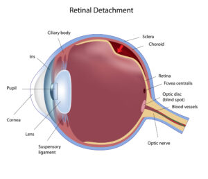
Retinal detachment is an eye condition where the retina, found in the back of the eye, detaches from the surrounding tissue. The retina has light-sensitive cells responsible for converting light into neural impulses.
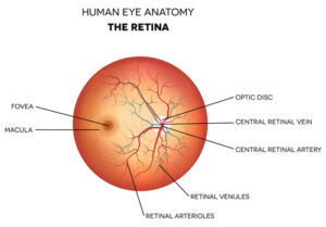
These impulses communicate with your optic nerve and brain, allowing them to process what you’re seeing. When the retina separates from the supportive tissue, it no longer receives the nourishment it requires.
Consequently, your eyes may experience irreversible damage in a short period. Your eye doctor will conduct a detailed eye exam by dilating your pupil to diagnose a retinal detachment. Continue below to learn more about retinal detachments and how they can seriously impact your vision!
Types of Retinal Detachment
There are three main kinds of retinal detachments. They are:
Exudative
An exudative retinal detachment is usually caused by eye trauma, inflammatory disorders, and other retinal diseases. In this type of detachment, fluid leaks under the retina even without breaks or tears in the retina.
Rhegmatogenous
A rhegmatogenous retinal detachment happens when a tear in the retina enables fluid to get under it. When fluid accumulates under the retina, it separates it from the cell layer that provides nourishment.
Tractional
With a tractional retinal detachment, scar tissue on the surface of the retina begins to contract. This scar tissue contracting forces the retina to detach from the retinal pigment epithelium.
Risk Factors for a Detached Retina
Factors that increase the risk of a retinal detachment include:

- Most people that experience a retinal detachment are over 40 years old but you can have one at any age
- If you’ve had a detachment in one eye, it increases your risk of developing it in your other eye
- Blunt force eye trauma
- Thinning of your peripheral retina
- Patients with diabetes
- Severe nearsightedness
- Having a previous procedure like having cataract surgery
- Family history of retinal detachment
- Eye disorders and diseases like degenerative myopia, retinoschisis, and uveitis
- Retinal tear
Signs and Symptoms of Retinal Detachment
- Blurred vision
- Flashes of light
- Feeling as though a curtain is closing in on your peripheral vision
- Sudden onset of multiple floaters. These are tiny specks that appear to move in your field of vision
Treating Retinal Detachment
Most patients with a detached retina will require surgery to put the retina back in its correct position. Fortunately, 90 percent of retinal detachment treatments succeed after only one procedure.
Most patients undergo a surgical procedure to treat their retinal detachment within a day or two of receiving a diagnosis, especially if central vision and macula aren’t affected yet.
If you need to repair a retinal detachment, three options are available depending on the type of detachment. Your surgeon will discuss the treatment options best for you. They include the following:
Pneumatic Retinopexy
A pneumatic retinopexy is a suitable procedure if you have a small detachment due to a single tear at the top of the retina. It entails injecting a tiny gas bubble inside your eye to plug the tear that caused the detachment.
Plugging the tear enables the fluid to collect at the back of your retina, which gradually becomes reabsorbed. This way, the retina reattaches.
It takes a few days before the retina adheres properly. Once this occurs, your surgeon closes off the retinal hole using cryotherapy or laser photocoagulation. The bubble self-reabsorbs over several days.
Vitrectomy
During a vitrectomy, your surgeon creates three tiny incisions in the white part of your eye. Then, they use small instruments to guide and extract the vitreous fluid collected under your retina.
In its place, your surgeon inserts a silicone oil or a gas in the eye. Inserting the oil or gas pushes the retina to the wall of the eye. Then, to seal the retinal breaks, they’ll use laser photocoagulation.
Over several weeks, you’ll absorb the gas into your body. This enables the vitreous to refill your eye. However, if your surgeon used silicone oil, they will perform another procedure to remove it after a few weeks.
Scleral Buckling
With scleral buckling, your surgeon attaches a buckle or silicone band to the sclera over the tear. But before that, they will drain the fluid beneath the retina.
After draining the fluid that was under the retina, the surgeon sutures in the buckle. The scleral buckle stops the force pulling your retina outward, enabling it to go back to its usual position.
After securing the retina against the wall of the eye, your surgeon will use a laser to weld the edges of the retinal tear and seal the leak.
What Recovery is Like After Retinal Detachment Surgery

Once you’ve had surgery to fix a retinal detachment and tear, you’ll need to take time to recover. After your procedure, your eye doctor will prescribe eye drops to keep your pupils from closing or opening too widely.
In addition, they may give you an eye shield or patch to wear over your eye. You’ll only have to wear this for a few days.
While recovering, you might experience:
- Blurry vision
- Eye pain
- Red, swollen, or tender eyes
If the surgeon uses a gas bubble to flatten your retina, they’ll advise you to make sure your head is in a particular position for a certain number of days. After that, it will take about two to four weeks before you can resume your daily routine.
Remember, follow-up care is a vital part of your complete recovery and safety. Make sure you go to any scheduled follow-up appointments.
However, you should contact your eye doctor immediately if you have any of the following signs or symptoms:
- Thick discharge from your eye
- A fever
- Sudden shortness of breath or chest pain
- Abrupt vision changes
- Severe eye pain
- Experiencing new flashes of light
Retinal detachment is a vision-threatening condition that needs prompt treatment by a specialist. Our experienced, board-certified ophthalmologists at Evergreen Eye Center are experts and talented professionals in diagnosing retinal detachment.
Have questions or concerns when it comes to retinal detachment? Request an appointment at Evergreen Eye Center in Tacoma, WA, now!
